Introduction
Prof. Toshiyuki Oda
In ancient Greece, Aristotle believed that living organisms were composed of three hierarchical levels. The first level consisted of the unorganized physical substances, made up of the four elements: earth, water, air, and fire. The second level was the homogeneous parts, or tissues, and the third level was the heterogeneous parts, or organs. Until the cell theory was proposed by Schwann in 1839, biologists regarded anything smaller than tissues as little more than an amorphous, inorganic substrate. Today, with the advancements in molecular biology, we know that the cytoplasm is far from being just a formless substrate. It is a dynamic environment where tiny molecules, each a millionth of a millimeter, actively move around, sustaining life, including that of humans. In our laboratory, we harness cutting-edge technologies, such as cryo-electron microscopy, to explore this nanoscale world within cells, aiming to unravel the mysteries of life.
Oda’s group:Cryo-electron “histology”
Cryo-electron microscopy (cryo-EM) is an advanced technique that allows us to observe samples in a cryogenic environment. This method preserves biomolecules in their natural state while providing extremely high-resolution images. Unlike traditional methods, cryo-EM enables detailed analysis of the three-dimensional structures of biomolecules, making it a widely utilized tool in life sciences today.
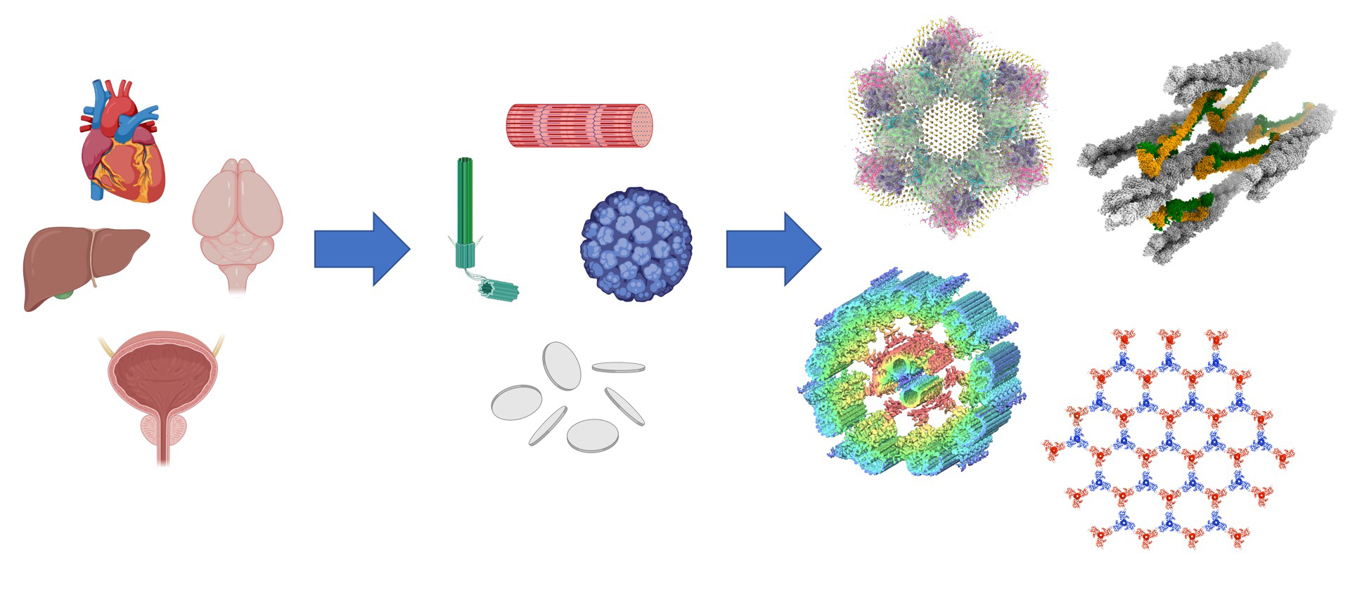
At the core of this research theme is our pioneering approach known as “cryo-electron histology.” This cutting-edge technique uses cryo-EM to uncover the intricate architecture of protein complexes and organelles unique to specific tissues such as the heart, skin, and bladder. By focusing on these complex structures, we aim to bridge the fields of anatomy and structural biology, ultimately achieving a deeper understanding of the microscopic architecture within living organisms.
Exploring the Diversity of Mysterious Marine Animals – Placozoans
Assistant professor Munetsugu Bam
Placozoans are among the earliest diverging multicellular animals, characterized by their simple body structure that lacks muscles, nerves, and circulatory systems. Despite their simplicity, these animals exhibit fascinating and unexplained phenomena, such as maturing sexually and forming eggs and yolk within their bodies (though development to maturity has not been observed in laboratory cultures). Additionally, placozoans are colorless and transparent, making them difficult to observe directly in the field, and little is known about their natural ecology.To uncover the mysteries of these enigmatic animals, I conduct both laboratory experiments using cultured specimens and fieldwork investigations.
In addition to research on placozoans, I am also involved in marine education activities in Yamanashi Prefecture, a landlocked area, using marine organisms. I also conduct ecological surveys of the purple sea urchin (Heliocidaris crassispina) at Mochimune Beach in Shizuoka City and research on sea urchin aquaculture using discarded vegetables in collaboration with Pickles Corporation and Yaizu Fisheries High School.
-scaled.jpg)
Takahashi’s group:Mechanism of Individuality in Neural Activity
Assistant Professor Hironori Takahashi
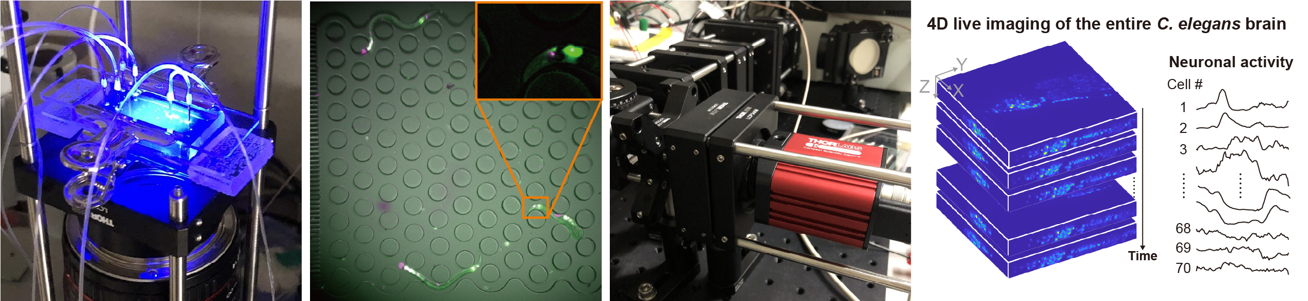
Animals, including humans, exhibit a variety of responses to the same sensory stimuli. It remains unclear whether such individual differences are merely noise in neural information processing or an essential function for ensuring diversity. In Autism Spectrum Disorder (ASD), these individual differences are significantly pronounced, making the elucidation of their mechanisms a critical issue from a disease perspective. Our laboratory aims to uncover the neural mechanisms underlying individual differences using the small nematode Caenorhabditis elegans (C. elegans) as a model organism. With its compact brain model consisting of about 300 neurons, we employ the following experimental techniques in our research:
Whole-brain imaging / High-throughput neural imaging /Molecular biology techniques such as construct creation / Mathematical analysis of neural activity data / Analysis using machine learning / Various image analysis methods / Development of original tools through programming /Synapse observation using electron microscopy